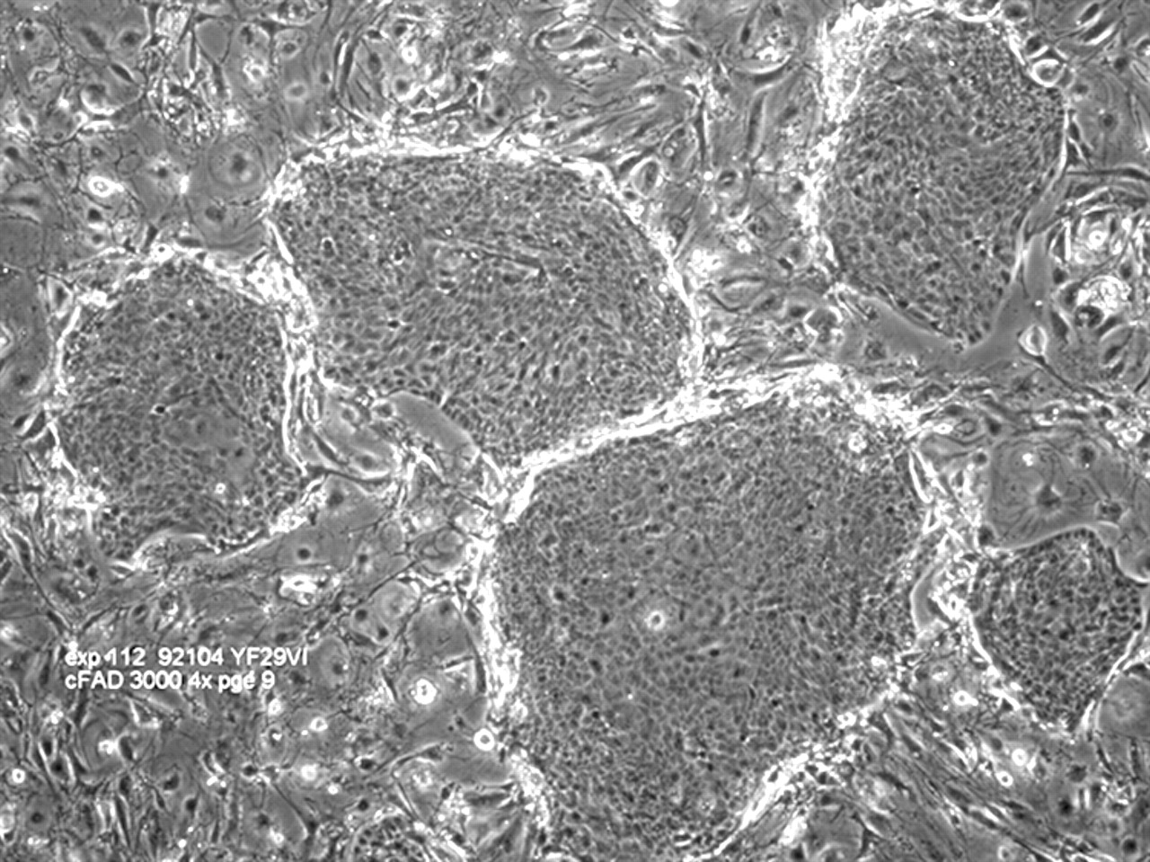
Human Keratinocyte Cultures as Models of Cutaneous Esterase Activity
As the quest to develop useful in vitro test methodologies to replace rabbit eye and skin irritation tests continues, validation projects still depend on historical rabbit eye and skin irritation data as the benchmark against which to measure the performance of the in vitro assays.
During our daily contact with sponsors, we are asked more and more about the relationship between in vitro test methods and the results of human trials, especially in the area of skin irritancy.
To date, there is a limited amount of data available in the literature which compare the results of in vitro methodologies with those of human volunteer trials. In this study, data obtained in a simple cytotoxicity assay, the 3T3 neutral red uptake (3T3 NRU) assay, will be compared with data from a standard three-application patch test.
A selection of 20 personal care and cosmetic products will be tested in a single-blind, within-subject comparison three-application patch tests on a panel of 30 healthy human volunteers.
Skin irritation potential will be determined by assessing and scoring the volunteers’ reactions to the products.
Products will be ranked according to their irritancy potential in vivo and in vitro, and the relationship between each of the EC values of the NRU assay and patch test results will be determined.
A reproducible, quantifiable assay has been developed for the measurement of esterase activity in human keratinocyte culture, using the model substrate 4-methyl umbelliferyl heptanoate (MUH) which is hydrolysed to the fluorescent metabolite 4-methyl umbelliferone (MU).
Activity was assessed in two human keratinocyte cell lines, NCTC 2544 and SVK-14, and in freshly isolated human breast keratinocytes from primary culture to passage 3. Vmaxvalue for MUH hydrolysing activity in the two cell lines showed that the less differentiated cell line NCTC 2544 (Vmax = 23.00 ± 2.84) expresses a much higher activity than SVK-14s (Vmax = 13.28 ± 1.42) which are more differentiated and able to form a cornified envelope.
Activity in the freshly isolated human breast keratinocytes decreased with time in culture in all three donors tested, which is also likely to relate to the extent of cell differentiation.
In human skin, xenobiotic esters penetrating the stratum corneum may be exposed to changing levels of hydrolysing esterases as they are absorbed across the epidermal cell layers.
The assay for MUH hydrolysis will be a useful tool for the study of esterase activity in populations of human keratinocytes in vitro.
“…Use of collagen, both native and modified, as a substrata for growth of cells from different tissues has been reported extensively [4,5,6,7,9,17,23].
In the present investigation, we have employed sources of collagen that are available in relatively pure form and have also not been extensively investigated earlier.
The serosal layer of bovine intestines and bovine Achilles tendon are very pure sources of bovine collagen and are available as byproducts of the slaughtered animals.
This study is aimed at the evaluation of wound dressings prepared from these two sources and cross-linked with agents 1,6-diisocyanatohexane (HDI) and basic chromium sulfate (BCS), for clinical use…”
